

Thermography
INFRARED THERMOGRAPHY
Definition
Infrared thermography is the science of capturing and analyzing thermal information using thermal imaging devices from a distance. The thermal camera works by detecting infrared radiation from the surface of a body that is invisible to the human eye and converting it into an image that we can see.
Therefore, thermal images represent levels of radiation intensity from the surface of the horse’s skin, thus detecting changes in dermal blood flow.
Application in Veterinary Medicine
Infrared thermography is a complementary method of diagnosing musculoskeletal and nervous system diseases. With this examination, the timely detection of subclinical orthopedic problems is feasible, which is important for their rapid and effective management.
Infrared thermography can identify injured areas of the body (e.g., tendons) 2 to 6 weeks before they become clinically apparent (lameness, swelling, etc.).
An experienced and certified thermographer, using specialized software, can analyze thermal images of the horse and assist in the diagnosis of orthopaedic problems, as well as provide useful information to the horse owner or trainer. Special care is needed, as the inappropriate or unprocessed thermal images may lead to misinterpretation of the results.
The examination is carried out under specific conditions and takes only a few minutes. After the examination is completed, and if the owner wishes so, a report can be drawn up in writing, presenting the thermal images and the results of their analysis.
We have internationally recognized certification in Infrared Thermography (ITC Cat.1 Thermographer).
WHICH CONDITIONS CAN BE DETECTED WITH THE USE OF THERMAL CAMERA?
Examples of situations where the use of Infrared Thermography is applicable:
- Tendon and ligament injuries
- Impingement of the dorsal spinous processes (‘Kissing Spine’) – Figure 1
- Evaluation of saddle fit – Figure 2
- Hoof abnormalities
- Arthritis
- Cervical problems
- Subclinical inflammations that may affect athletic performance
- Pre-purchase examination
THERMAL IMAGE EXAMPLES
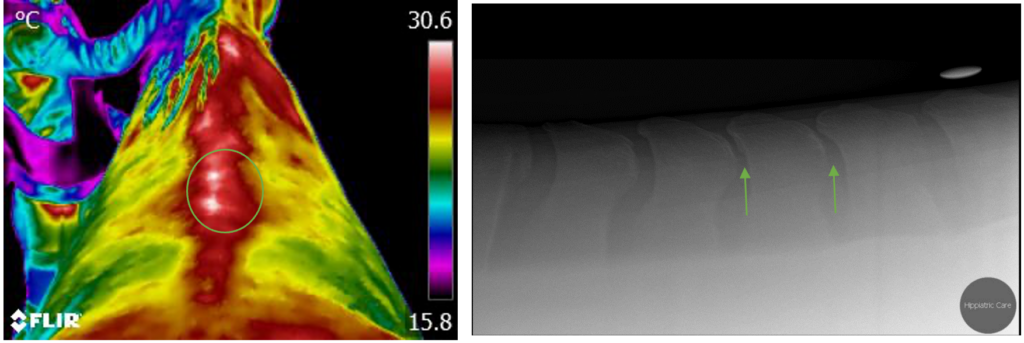
Figure 1: The thermal images were first taken, and then x-rays showed crowding of the interspinous spaces at two locations in the caudal thoracic spine.
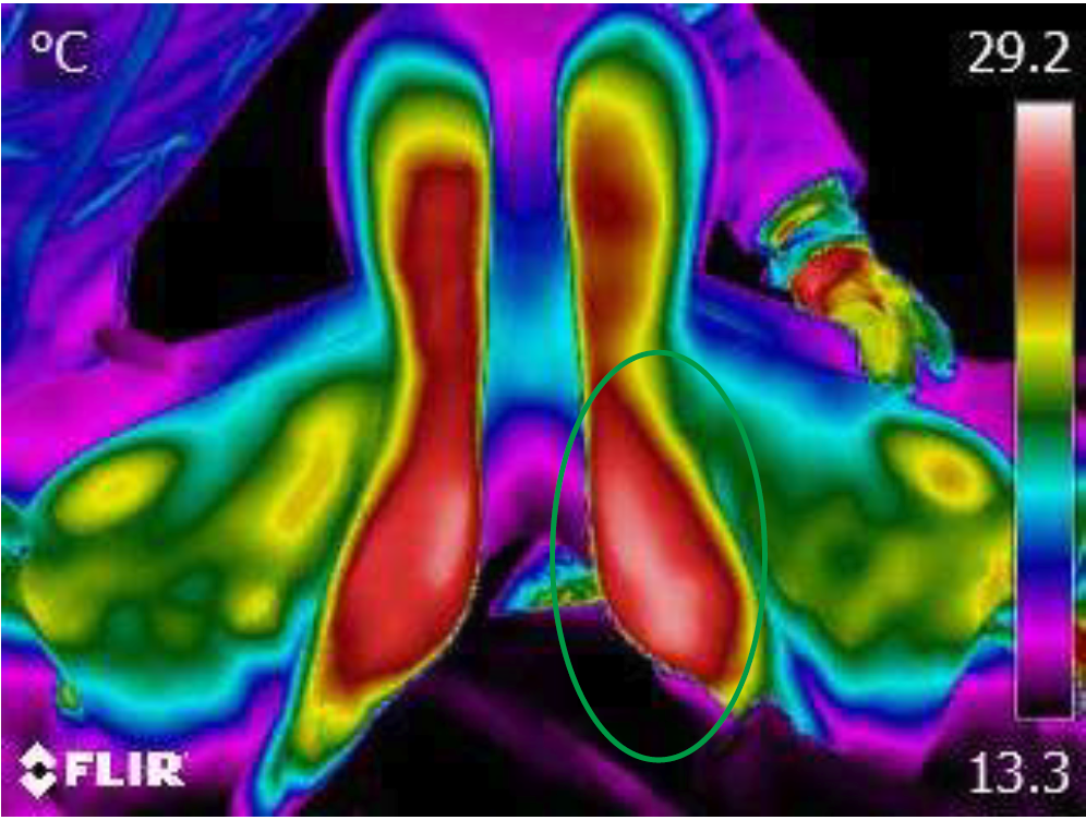
Figure 2: Thermographic image of the saddle to detect areas that receive more pressure.
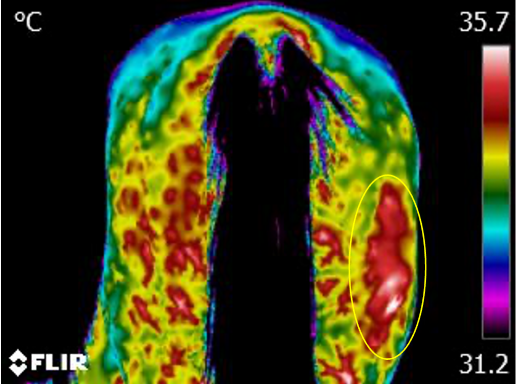
Detection of an inflamed area that developed into an abscess (circle).
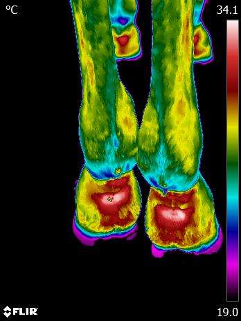
Thermographic image for the detection of signs of inflammation in the hind limbs.